 Skip to content
Pelvis & Pelvic floor Anatomy
Pelvic floor is a super trendy expression with new exercises to do or machine to train them. We use it more and more as we realised how it is important for women to protect and to exercise it correctly. But do you actually know what is it exactly? Do you have an idea how it looks like? No? So today let’s have a look at Pelvis & Pelvic floor Anatomy in order to understand how it works and to then be able to protect and exercise it correctly!Pelvis bones
It is a complex of bone enclosing visceral organ of the perineum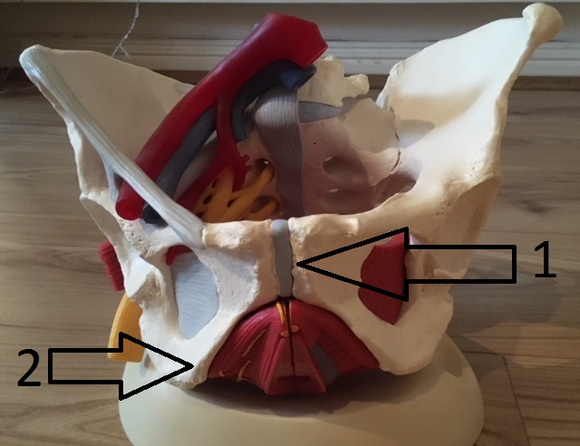
- #1: Pubis symphysis is a junction (joint) between two bones. In pregnancy a lot of pressure is applied on this junction and may lead to inflammation and pain in this area or spread lower through the adductor muscles which insert directly on the Pubis symphysis
- #2: Ischions are the bony prominence on which we sat. In pregnancy, this area is often painful due to muscles spasm of the piriform (here a video to stretch it)
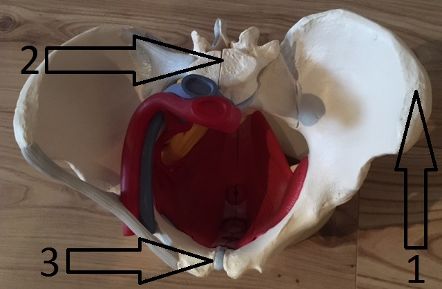
- #1 : Iliac crest are the bony prominences you can feel on the side of your lower abdomen
- #2 : vertebrae
- #3: Pubis symphysis
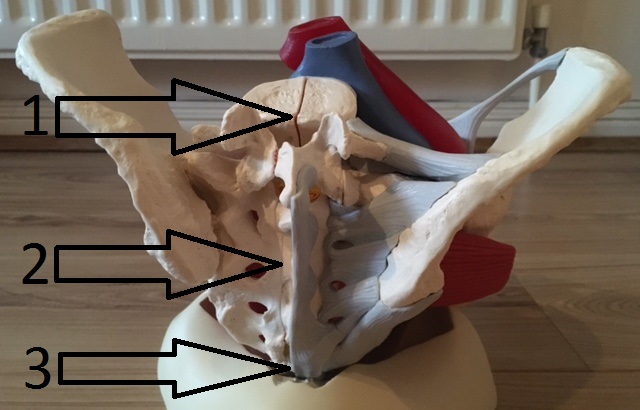
- #1 Vertebrae
- #2 Sacrum is a big bone at the end of your spine. It ends with a small bone coccyx!
- #3 Coccyx
Genital organ
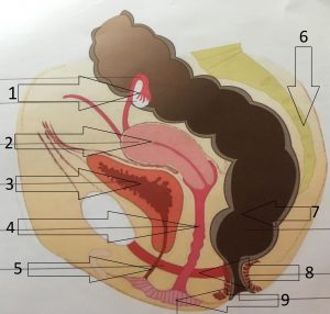
- #1 Ovary
- #2 Uterus is held by ligaments attached to the sacrum
- #3 Bladder rest on the pubis symphysis
- #4 Vagina is a big gap in the middle of the pelvis
- #5 Urethra is a canal to empty the bladder
- #6 Sacrum
- #7 Rectum is terminated by an angle and the anus to retain faeces allowing anal continence
- #8 Pelvic floor muscles
- #9 Vaginal opening
Pelvic Floor muscles
These muscles form a hammock holding the pelvis organs (bladder, uterus, vagina, rectum…) in place.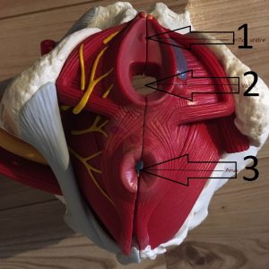
- #1 Urethral orifice
- #2 Vagina
- #3 Anus
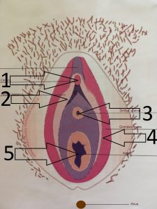
- 1# Clitoris
- 2# Labia minora
- 3# Urethral Orifice
- 4# Labia majora
- 5# Vagina
Now let’s see the muscles hiding behind, the pelvic floor muscles! There are 3 different layers: superficial, intermediate and deep muscles.
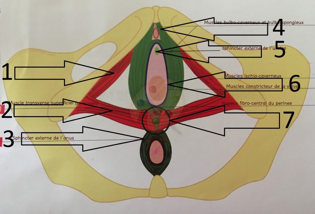
- 1# Ischiocavernosus muscle
- 2# Superficial transverse perineal muscle helps to close the urethra and allow urine continence
- 3# External anal sphincter as suggest by it is name, it close the anus 😉
- 4# Bulbospongiosus muscle inserts around the clitoris. It helps to erect it and to close the vaginal opening
- 5# External urethral sphincter as its name say, it close the urethra and allow urine continence
- 6# Bulbo coccygeal muscle is an involuntary muscle and also helps to close the vaginal opening. It is always tear during natural birth
- 7# Fibrous Perineal body
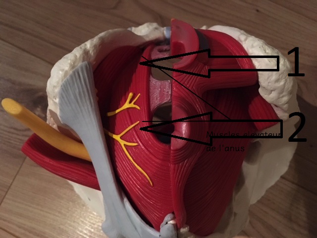
- #1 & #2 Levator ani muscles allow anus contraction and anal continence but also trigger its relaxation allowing defecation.


3 Responses
Great layout! And the bulbo coccygeal muscle doesnt always tear during a natural vaginal birth. Sure, sometimes there are tears, but not always, and some tears occur elsewhere.
Thanks for this great article!
You’re welcome and thank you for your comment 🙂
Wish there had been such an informative site when I was having pregnancies however I will now use the exercises which should help slight urinary urgency. Many thanks for the site it should help many